|
||
|
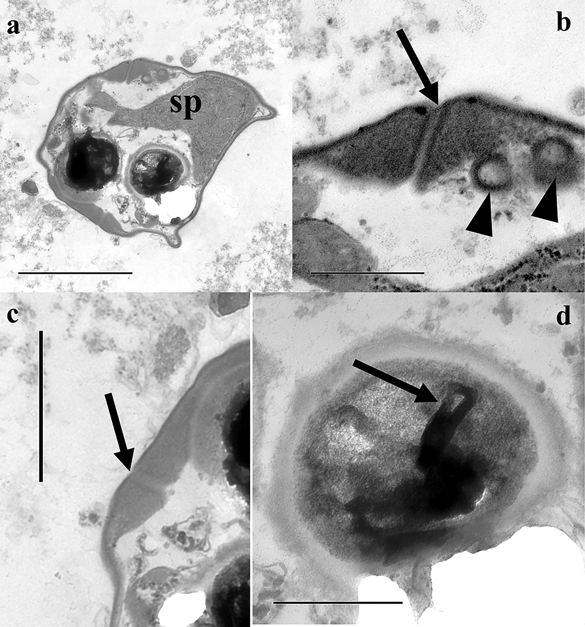
Transmission electron microscopy images of Ceratomyxa amazonensis isolated of Symphysodon discus from the Unini River, Amazonas State, Brazil. a. Myxospore showing two sub-spherical polar capsules and sporoplasm (sp) occupying most of the myxospore volume; b. Detail of the apical suture (black arrow) and sporoplasmosomes (arrowheads); c. Detail of lateral suture (black arrow); d. Polar capsule displaying still uncoiled internal polar tubule (black arrow). Scale bars: 2 µm (a); 1 µm (c); 500 nm (b, d). |