|
||
|
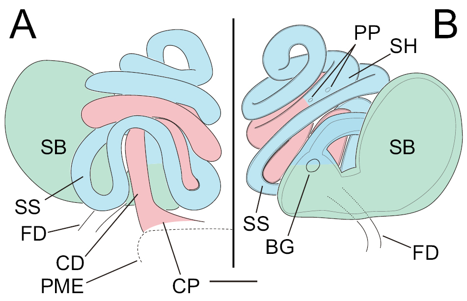
Cybaeus melanoparvus Kobayashi, schematic drawing of epigyne and spermathecae, based on female specimen (KUZ Z3765). A. Ventral view; CD, copulatory duct; CP, copulatory pore; FD, fertilization duct; PME, posterior margin of epigynal plate; SB, spermathecal base; SH, spermathecal head; B. Dorsal view; BG, Bennett’s gland; FD, fertilization duct; PP, primary pore; SB, spermathecal base; SS, spermathecal stalk; SS, spermathecal stalk. Duct from CP to before PP colored in red; duct from PP to before SB colored in blue; SB colored in green. Scale bar: 50 µm. |