|
||
|
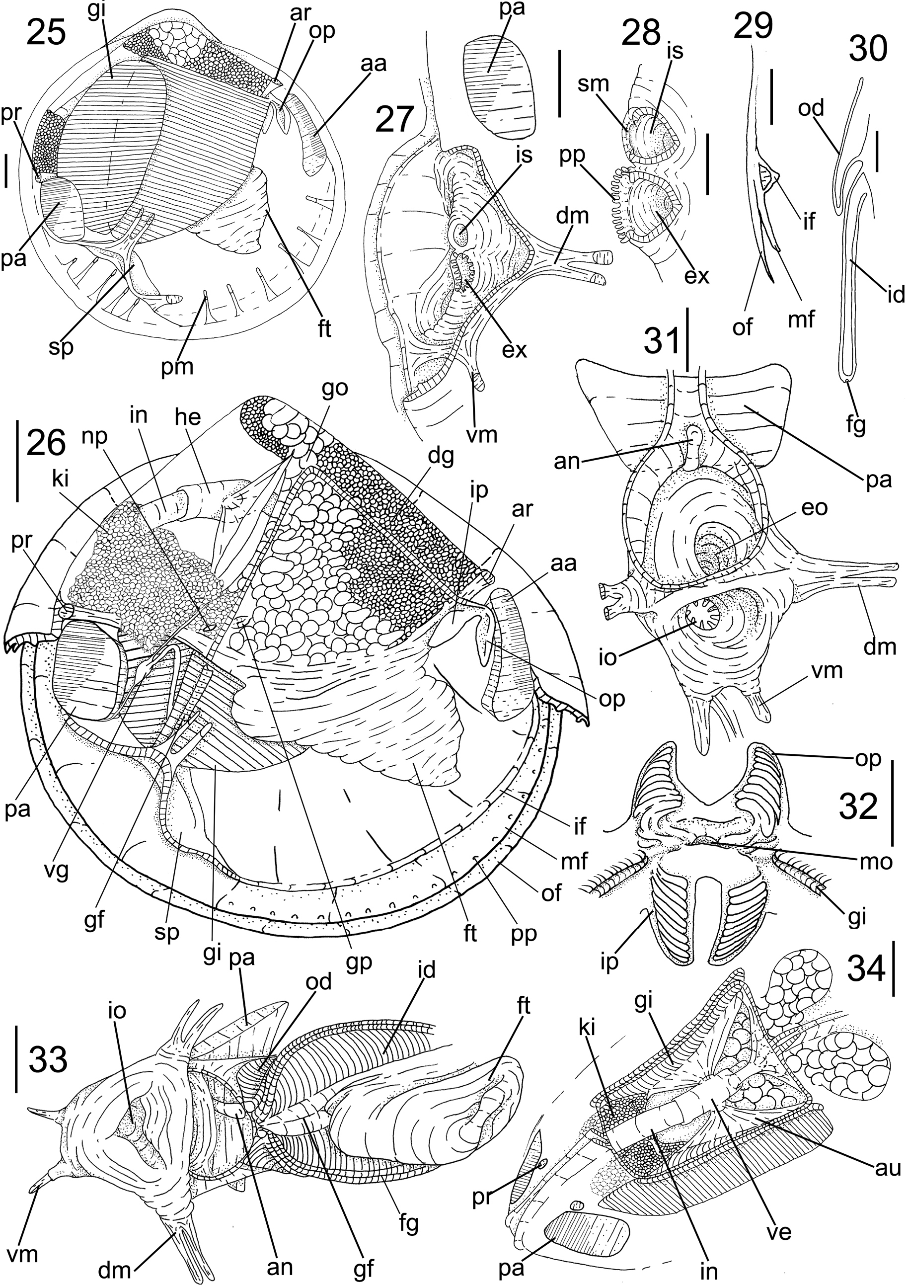
Anatomy of Cyrenoida floridana (FMNH 328260). 25. Right view valve removed, some structures seen by transparency of mantle lobe; 26. Same, with mantle and gill removed; 27. Incurrent and excurrent siphons, posterolateral view, right mantle lobe partially removed, some adjacent structures shown; 28. Siphonal tips; right view, both partially sectioned longitudinally; 29. Mantle border, section in its ventromedial portion; 30. Gill, transverse section in its central portion; 31. Incurrent and excurrent siphons, interior view, with details of their base and siphonal muscles; 32. Labial palps, ventral view, outer hemipalps deflected dorsally; 33. Posteroventral visceral region, ventral view, showing fusion of inner demibranchs in siphonal base; 34. Pericardial region, posterodorsal view, dorsal mantle wall partially removed. Scale bars: 2 mm (25–28, 31, 33–34); 1 mm (29, 30, 32). |