|
||
|
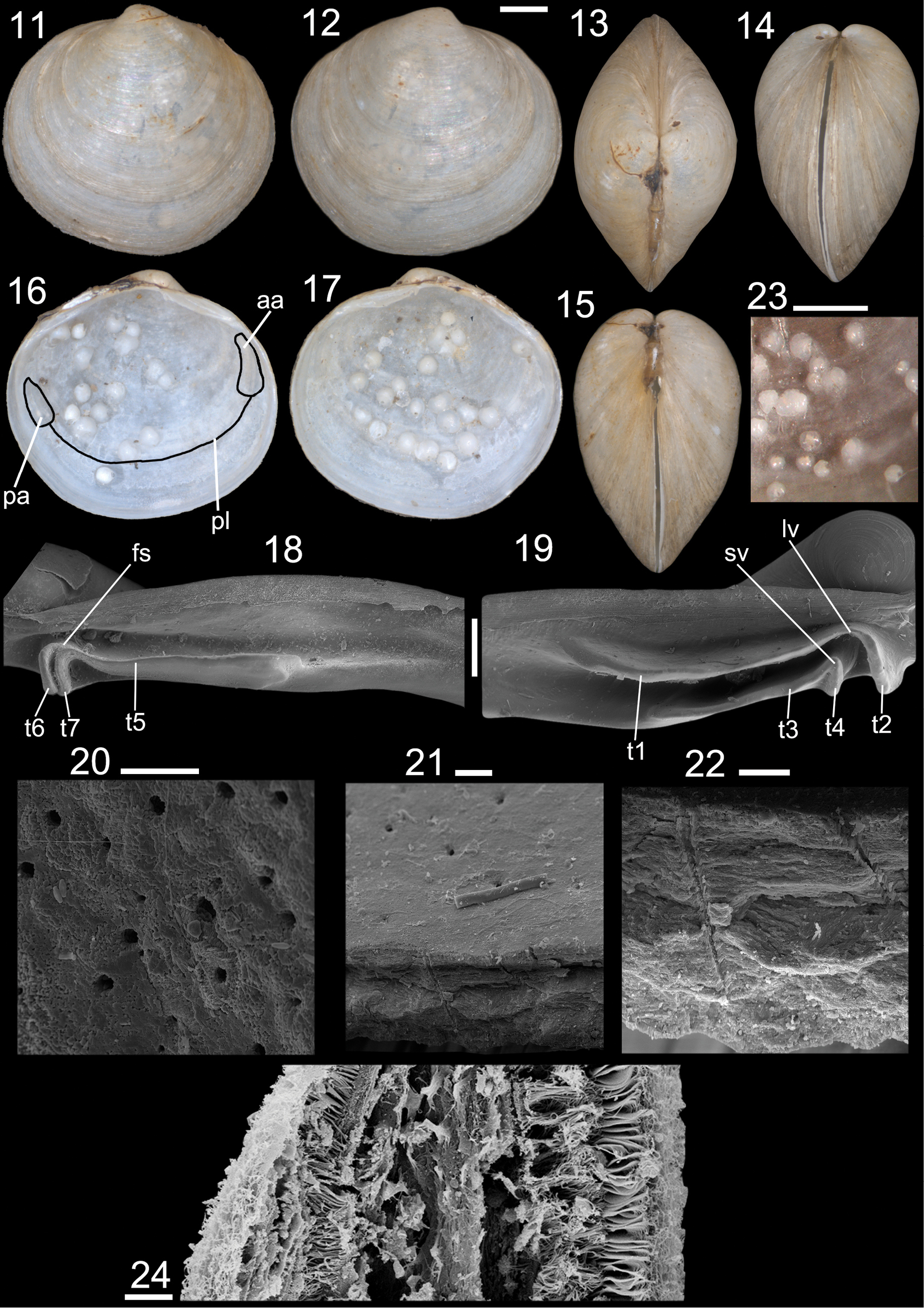
Cyrenoida floridana, shell and gills details. 11–22, 23. UF 246126; 24. FMNH 328260. 11. Left valve, external view; 12. Right valve, external view; 13. Dorsal view of shell; 14. Anterior view of shell; 15. Posterior view of shell; 16. Left valve, internal view, muscle scars and pallial line outlined; 17. Right valve, internal view; 18. Left hinge, SEM; 19. Right hinge, SEM; 20. Internal surface of shell, SEM, showing microtubule orifices; 21. Detail of microtubule patch; 22. Fractured shell showing microtubules partially through shell thickness; 23. Detail of nodules at shell internal surface; 24. Gill fragment, transverse section, SEM. Scale bars: 1 mm (11–17, 23); 200 µm (18, 19); 20 µm (20–22), 0.5 mm (23); 10 µm (24). |