|
||
|
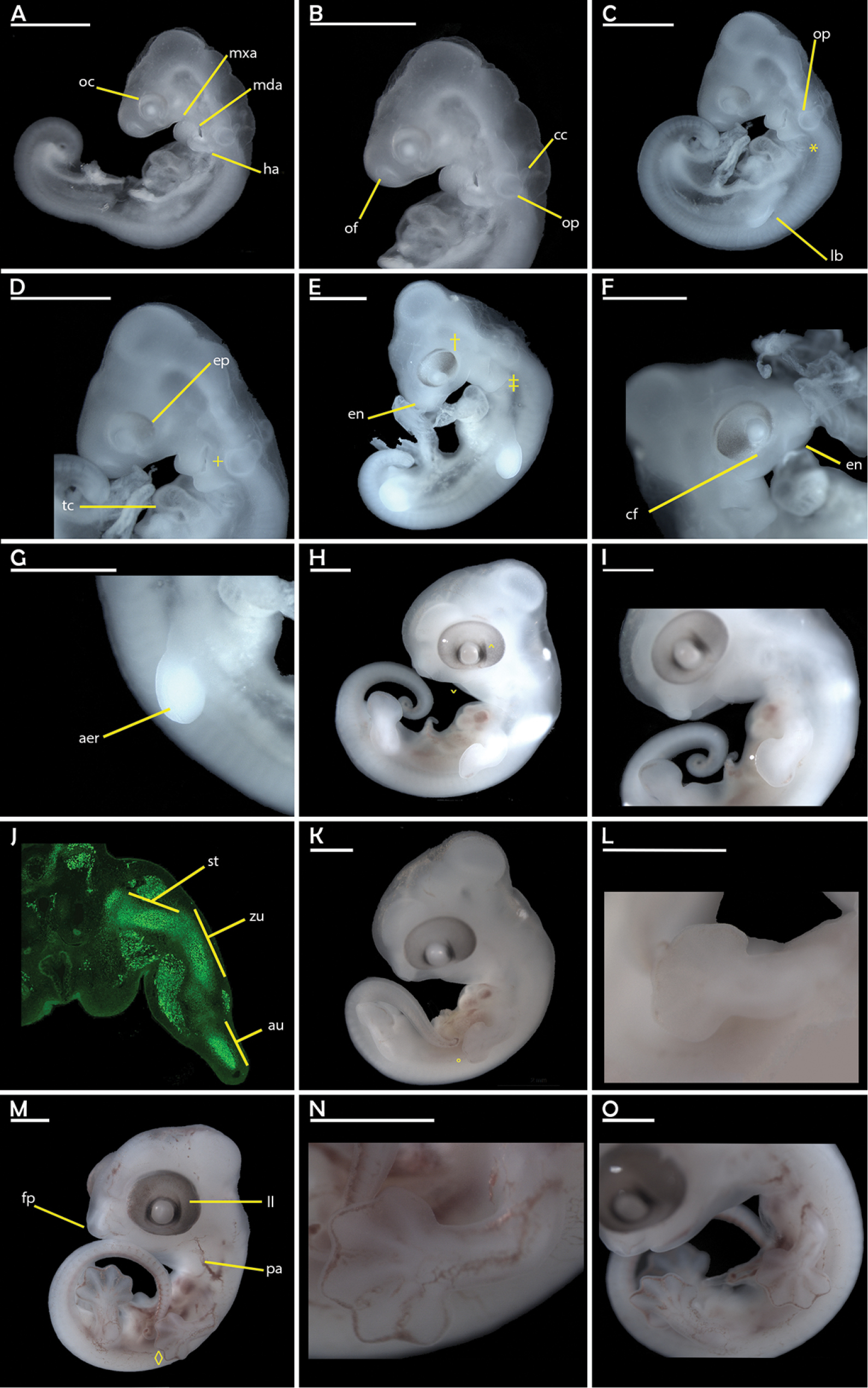
Overview of embryonic developmental stages A to F, erected in this study. Stage A is depicted in (A, B) and represented by CA2017_007. (C, D) show the representative CA2017_003 for stage B. Stage C (E–G) is represented by CA2017_006. CA2017_005 represents stage D in (H–J). Note the immunohistochemistry for SOX9 expression in a limb cross section in (J). Stage E is shown in (K–L), represented by CA2017_010. CA2017_012 represents stage F in (M-O). Except for (J), all pictures in lateral view. Scale bars equal 1mm. Abbreviations: aer – apical ectodermal ridge; au – autopod; cc – cartilage capsule; cf – choroid fissure; en – external nares; ep – eye pigmentation; fp – frontal nasal process; ha – hyoid arch; lb – limb bud; ll – lateral lines; mda – mandibular arch; mxa – maxillary process; oc – optic cup; of – olfactory pit; op – otic pit; pa – pharyngeal arches; st – stylopod; tc – thorac cavity; zu – zeugopod. |