|
||
|
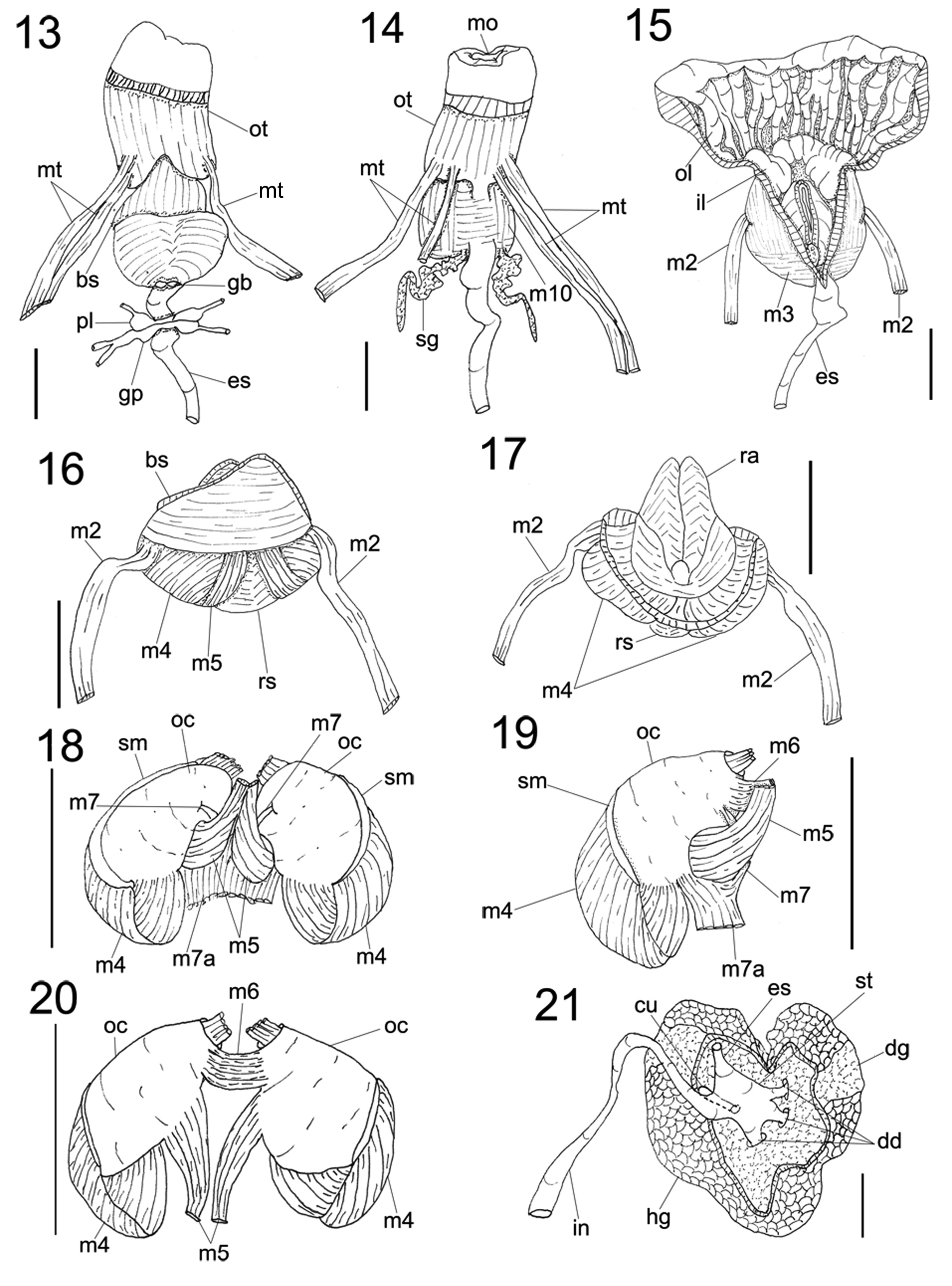
Actinocyclus verrucosus details of digestive system. 13. Foregut and nerve ring, ventral view, some adjacent structures also shown as in situ, scale: 1 mm. 14. Same dorsal view, scale: 1 mm. 15. Same, longitudinally sectioned, showing internal oral tube, dorsal view, scale: 1 mm. 16–20. Odontophore anatomy, scales: 1 mm. 16. Whole ventral view, sphincter removed. 17. Whole dorsal view, esophagus removed. 18. Dorsal view, radula removed, each cartilage slightly deflected. 19. Whole right view. 20. Ventral view, m4 and m5 folded down to expose odontophore cartilage; m7 and m7a removed. 21. Midgut as in situ, dorsal view, scale: 2 mm. |