|
||
|
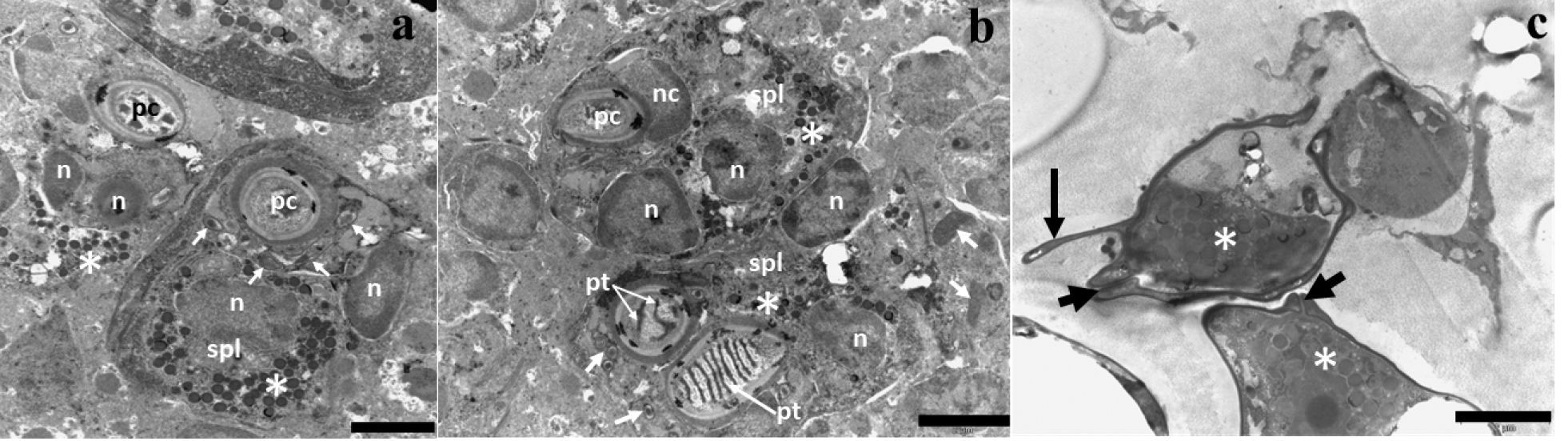
Internal ultrastructure by TEM of myxospore of Henneguya sp. infecting skin of Synbranchus marmoratus. a: sporoblast in young developmental stage showing binucleated sporoplasm (n) contained several sporoplasmosomes (asterisk), valve-forming materials (white arrow) and polar capsules (pc) with absence of polar tubule. b: polar capsule (pc) with capsular nuclei, polar tubule internalized contained seven to eight coils (pt), sporoplasm binucleated (spl/n) and contained sporoplasmosomes (asterisk) at a more advanced sporoblast developmental stage. c: Spores with sutural lines (small arrows), sporoplasm with numerous sporoplasmosomes (asterisk) and caudal appendage (large arrow). Scale bars: 2 µm. |