|
||
|
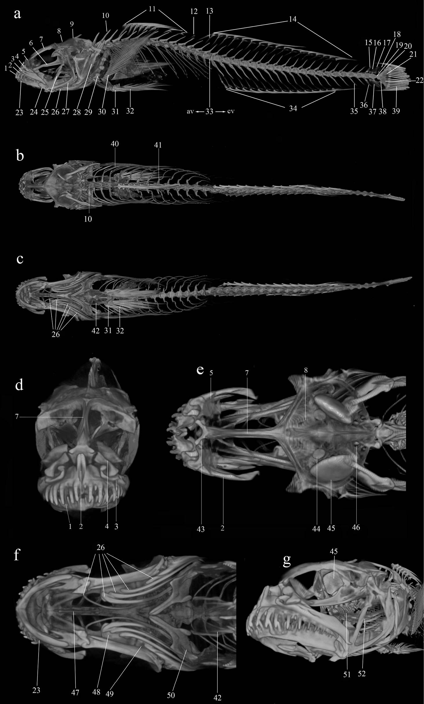
Micro-CT images of right (a), dorsal (b), and ventral (c) views of specimen YSFRI27216; front (d) dorsal (e), ventral (f), and oblique view (g) of the head of specimen YSFRI27216. 1. teeth, 2. premaxilla, 3. maxilla, 4. palatine, 5. ectethmoid, 6. parasphenoid, 7. frontal, 8. parietal, 9. supraoccipital, 10. neural spine, 11. first dorsal fin spines, 12. interdorsal pterygiophores, 13. pterygiophore, 14. second dorsal fin rays, 15. neural spine of preural centrum 3(NPU3), 16. neural spine of preural centrum 2(NPU2), 17. epural 1 (EP1), 18. epural 2 (EP2), 19. urostyle, 20. hypural 5 (HY5), 21. hypural 3+4 (HY3+4), 22. hypural 1+2 (HY1+2), 23. dental, 24. articular, 25. sympletic, 26. branchiostegal rays, 27. preopercular, 28. subopercular, 29. proximal radials, 30. pectoral fin soft rays, 31. pelvic fin spine, 32. vertebral canal, 33. boundary of abdominal vertebra and candal vertebrae, 34. anal fin rays, 35. ventrispinales, 36. haemal spine of preural centrum 3 (HPU 3), 37. haemal spine of preural centrum 2 (HPU 2), 38. parhypural (PH), 39. caudal fin ray, 40. rib, 41. parapophysis, 42. pelvic bone, 43. ethmoid, 44. lapillus 3+4 (HY3+4), 45. sagittae, 46. asteriscus, 47. basihyal, 48. ceratohyal, 3 (HPU 3). 49. epihyal, 50. cleithrum, 51. pharyngeal tooth, 52. ceratobranchial. |