|
||
|
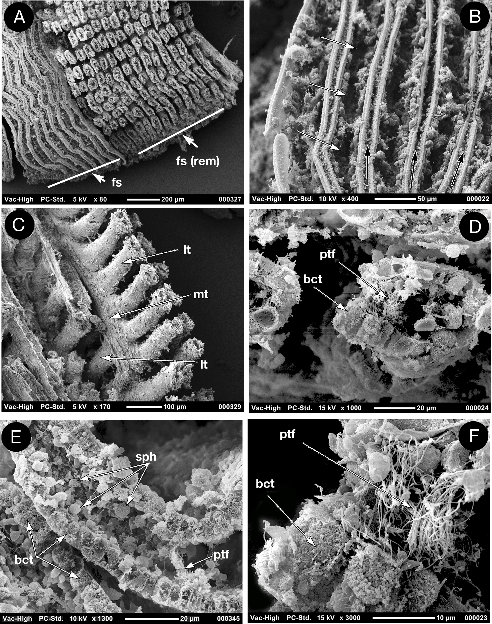
Scanning electron micrographs of the ctenidium of Ochetoctena tomasi, A frontal face, frontal surface intact on the left, removed on the right to reveal tubules; B frontal surface with entrances to tubules arrowed; C single filament showing lateral and median tubules; D cross section of a single tubule; E long section of adjacent tubules; F bacterial bundles within bacteriocytes and paddle tipped filaments in the lumen of the tubule. bct bacteriocyte; spb spherical body; ptf paddle tipped filaments; fs frontal surface; fs (rem) frontal surface removed; lt lateral tubule; mt median tubule. |